Echocardiography
This test can also be called a transthoracic echocardiogram, but is usually referred to simply as an echocardiogram or echo for short.
What is an echocardiogram?
An echocardiogram, also known as an echo, is a test that uses sound waves to build up a moving picture of the heart. It is similar to the ultrasound scan used in pregnancy and is extremely safe. The ultrasound waves reflect against structures in the heart to measure the size and function of your heart and to check how well your heart is working. An echo can also show the direction and velocity of blood flowing through your heart and across your heart valves.
How is an echocardiogram performed?
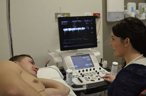
A doctor or cardiac technologist will perform the echo. We will ask you to remove your clothes from the waist up and to lie on an echocardiogram bed on your back. We may ask you to roll over onto your left-hand side during the test.
Three small sticky patches, called electrodes, will be attached to your chest and connected to the echo machine. We will place a small hand-held transducer (echo recorder) on your chest. This will have a small amount of lubricating jelly on it. Images of your heart will then be taken from different positions on the chest. The recorder sends high-frequency sound waves to the heart and records the echoes of sound waves reflected back from your heart. The echocardiography machine receives these echoes as electrical impulses and converts them into moving pictures of the heart.
Why do I need an echocardiogram?
An echo gives detailed images of your heart. It allows us to accurately assess the size of your heart, its chambers and how your heart valves are functioning. We can also examine the direction and velocity of blood flowing through your heart.
An echo allows doctors to diagnose, assess and regularly check:
- Heart murmurs
- Abnormal heart valves
- Atrial fibrillation
- Pump function in patients with damaged heart muscle (after heart attacks)
- Pump function of the heart for people with heart failure
- Infection on heart valves (infective endocarditis)
- Fluid collections around the heart (pericardial effusion)
- The source of a blood clot after stroke
- Congenital heart disease
- Pulmonary hypertension
How long does an echocardiogram take?
A short examination in an uncomplicated case may be performed in 15-20 minutes. The additional use of Doppler (a special part of the ultrasound examination that looks at blood direction and velocity) may add an extra 10-20 minutes. However, it may take up to an hour when there are multiple heart problems and the echocardiogram is more complicated.
When can I expect to receive the results?
A cardiologist will report on your echocardiogram. The results will then be sent to your cardiologist or doctor who will be able to discuss the results with you.